Look, contamination is the enemy. It’s bloody everywhere. Every substrate, every transfer, it’s an opportunity for unwanted organisms to move in. You gotta understand the biology or you’re just guessing (and losing batches).
Ecology of Contamination
Three factors matter. Just three.
Inoculum load. How many contaminant spores reach your substrate. Reduce it through sterile technique, air filtration, and environmental control.
Substrate susceptibility. How well the substrate supports contaminant growth. Nutrient density, pH and moisture, competing organisms, and how well you sterilised or pasteurised it.
Environmental conditions. Temperature, humidity, air movement. Many contaminants have different optima than cultivated species, which gives you opportunities to manage the environment in your favour.
Get any of these wrong and you’re done. Honestly, it’s that simple.
Fungal Contaminants
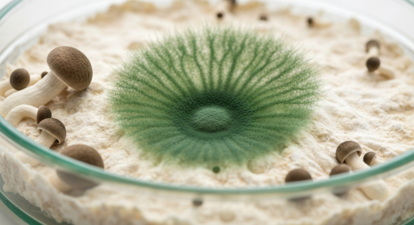
Trichoderma Species (Green Mould)
This is the big one. Most significant contaminant worldwide. We’re talking T. harzianum, T. viride, T. aggressivum, T. longibrachiatum. Ubiquitous soil fungus that’s an aggressive saprophyte and mycoparasite (it actively parasitises other fungi).
Key characteristics:
- Rapid growth. Grows 2-4x faster than cultivated species. On MEA at 25C, T. harzianum hits 15-25mm per day vs 3-8mm for Pleurotus ostreatus.
- Prolific sporulation. A single colony produces millions of spores dispersed by air currents.
- Mycoparasitism. Produces enzymes (chitinases, glucanases, proteases) that penetrate and digest other fungi’s hyphae. This isn’t just competition. It’s active predation.
- Broad tolerance. Grows 10-35C (optimum 25-30C), pH 2.0-8.0, moderate to near-saturation moisture.
- Antibiotic production. Produces trichodermin, gliotoxin, and peptaibols that inhibit mushroom growth.
Ever tried fighting this without bleach? Yeah, doesn’t go well.
Identification
| Stage | Appearance | Timeline |
|---|---|---|
| Initial colonisation | White mycelium, looks like target species | Days 3-7 |
| Sporulation onset | Green patches at periphery or stress points | Days 7-14 |
| Full sporulation | Dense bright green powdery masses | Days 10-21 |
| Advanced infection | Green with white advancing front, target retreating | Days 14+ |
Early Trichoderma is often misidentified as healthy growth. Key tell: it advances visibly faster and looks more cottony, less rhizomorphic.
T. aggressivum specifically targets Agaricus cultivation. Causes “green mould disease” that can destroy entire production cycles. Commercial farms use dedicated footwear, sanitisation stations, and HEPA filtration to keep it out.
Prevention
- Proper thermal processing. Conidia die above 60C, but if you recontaminate post-processing, it’s all for nothing.
- HEPA filtration (99.97% at 0.3um). Removes conidia (2-5um diameter).
- Environmental hygiene. Regular cleaning with 10% bleach. Spores accumulate on surfaces and in damp corners.
- Rapid colonisation. 15-20% spawn rate. Speed wins.
- pH management. 8.0-9.0 inhibits Trich while Pleurotus tolerates it fine.
Aspergillus (Black/Yellow/Green Moulds)
A. niger (black), A. flavus (yellow-green), A. fumigatus (grey-green). Thermotolerant. A. fumigatus grows optimally 37-42C. Xerotolerant (grows at water activity 0.75). A. flavus produces aflatoxins. Basically, don’t breathe this stuff in.
| Species | Colony Colour | Spore Size | Typical Source |
|---|---|---|---|
| A. niger | Black | 3.5-5.0um | Airborne, ubiquitous |
| A. flavus | Yellow-green | 3.0-6.0um | Grain, soil |
| A. fumigatus | Grey-green | 2.0-3.0um | Compost, soil |
Prevention: maintain substrate moisture at field capacity, adequate ventilation during incubation, store grain in cool dry conditions.
Penicillium (Blue-Green Mould)
Optimal 20-25C, pH 4.0-6.0. Moderate growth. Not mycoparasitic. Competes for resources through rapid colonisation and antibiotic production. If you see this, it indicates a sterile technique failure. Check your flow hood.
Prevention: improve sterile technique, HEPA or SAB for all open work, test inocula on agar before scaling up.
Cobweb Mould (Cladobotryum spp.)
Fruiting phase contaminant. Extremely rapid growth. Fine wispy transparent mycelium that drapes over the surface like a veil. Loves high humidity >90% RH and poor air circulation. Ever tried fruiting in a stagnant room? Yeah, cobweb loves that.
| Feature | Cobweb Mould | Healthy Mycelium |
|---|---|---|
| Texture | Fine, wispy, transparent | Dense, cottony or rhizomorphic |
| Colour | Grey-white | Bright white |
| Growth pattern | Spreading veil over surface | Penetrating into substrate |
| Growth rate | Very rapid (visible daily) | Moderate |
| Response to air movement | Slowed or halted | Unaffected |
Prevention and treatment: increase FAE (cobweb hates air movement), reduce humidity to 85-90% RH, spot treat with 3% hydrogen peroxide (kills cobweb on contact without harming cultivated species), remove severely affected substrates.
Lipstick Mould (Sporendonema purpurascens)
Pink to red-orange on grain. Diagnostic of under-sterilisation or excess moisture. Looks weird, smells bad. Prevention: dry grain properly before sterilisation, minimum 90 minutes at 15 PSI, review cooling procedures.
Neurospora (Orange Bread Mould)
FACILITY-LEVEL EMERGENCY. Billions of heat-resistant spores. A single contamination event can spread throughout your entire cultivation room. Seal and remove immediately. Deep clean entire facility with bleach. HEPA filtration of all incoming air. Prevention is far easier than remediation.
Bacterial Contaminants
Bacillus (Wet Spot / Sour Rot)
Most common bacterial contaminant in grain spawn. Forms endospores that survive boiling. D-value at 121C is 1.5-2.0 min. That sour smell? That’s the warning sign.
Presents as wet slimy sour-smelling grain 3-7 days post-inoculation. When you’re expecting white mycelium and instead see waterlogged, uncolonised grain.
| Root Cause | Mechanism | Prevention |
|---|---|---|
| Excess moisture in grain | Anaerobic zones protect endospores | Dry grain thoroughly, field capacity test |
| Insufficient sterilisation | Core doesn’t reach 121C | Extend time, vent cooker properly |
| Overloaded pressure cooker | Poor steam penetration | Reduce load, gaps between containers |
| Rapid cooling | Suck-back draws contaminated air in | Natural depressurisation only |
Pseudomonas (Bacterial Blotch)
P. tolaasii causes brown blotch on caps. Waterborne, spread by splashing. Simple fix, often ignored. Avoid splashing water on caps, maintain humidity without surface condensation, adequate air circulation, use chlorinated water 2-5ppm.
Environmental Risk Factors
| Parameter | Low Risk | Moderate Risk | High Risk |
|---|---|---|---|
| Temperature | 20-24C | 25-28C | >28C |
| RH (incubation) | 60-70% | 70-80% | >80% |
| Air filtration | HEPA (99.97% at 0.3um) | Pre-filtered (90% at 5um) | Unfiltered |
| Air changes (fruiting) | 4-8 ACH | 2-4 ACH | <2 ACH |
| Substrate moisture | 60-68% | 68-75% | >75% |
| Spawn rate | 15-20% | 10-15% | <10% |
| Time to spawning | <2 hours | 2-6 hours | >6 hours |
Each parameter interacts with the others. Perfectly pasteurised substrate spawned at low rate in a warm unfiltered room is still at high risk.
Contamination as Diagnostic Information
Every contaminated jar tells a story. Read it instead of just binning it.
| Observation | Likely Diagnosis |
|---|---|
| Green mould within 3 days | Substrate wasn’t sterilised properly |
| Green mould 7-14 days | Sterile technique failure |
| Bacterial wet spot in grain | Excess moisture, insufficient sterilisation, cold spots |
| Cobweb during fruiting | Poor air circulation, high humidity |
| Multiple contaminants in one batch | Facility hygiene issue |
| Contam near cooker wall | Uneven heat distribution |
| Contam limited to one inoculum | Contaminated culture, test on agar first |
Right. Know what you’re fighting, keep it clean, and move fast.
Related Reading
- Sterilization Methods. The primary defence against contamination
- Pasteurisation Techniques. How competitive exclusion manages contamination
- Substrate Chemistry. How chemistry influences contamination susceptibility
- Grain Substrates. Preparation protocols that minimise contamination risk